Tunica Adventitia is Continuous With Which Layer of the Heart
The blood vessels transport blood throughout the human body, They transport blood cells, nutrients, and oxygen to the tissues of the body, and they take waste and carbon dioxide away from the tissues, Blood vessels make up two closed systems of tubes that begin and end at the heart which are the pulmonary vessels and the systemic vessels.
Histology of the blood vessels
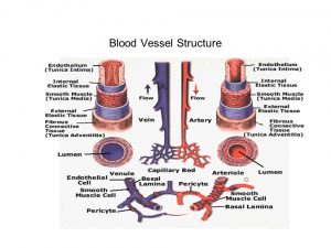
Histologically, the wall of any blood vessel, except capillaries, is formed of three concentric layers:
- Tunica intima, the innermost layer that faces the bloodstream, next to the lumen.
- Tunica media, the middle layer, its structure is related directly to the function of the vessel.
- Tunica adventitia, the outermost layer, it is usually continuous with the connective tissue of the surrounding structures.
Tunica Intima
It consists of:
- Vascular endothelium: simple squamous epithelial cells resting on a basement membrane. The cells are joined together by occluding junctions.
- Subendothelium: thin later of loose fibro-elastic connective tissue.
- In many arteries, the elastic fibers become condensed externally forming the internal elastic lamina.
Tunica media
It consists mainly of:
- Concentric layers of spirally-oriented smooth muscle fibers.
- The muscle fibers are separated by varying amounts of collagen, elastic fibers, and extracellular matrix of proteoglycans, all of which are produced by the vascular smooth muscle cells.
- Again in many arteries, elastic fibers may form the thin external elastic lamina separating the media from the adventitia.

Blood Vessel Structure
Tunica adventitia
It consists of:
- Longitudinally-oriented collagen and elastic fibers and fibroblasts.
- The adventitia also contains small blood vessels called vasa vasorum (vessels of the vessel), which provide nutrition to the vessel wall.
- It also contains nervi vasorum, which represents the vasomotor autonomic nerves.
Histological structure of arteries
Arteries are classified according to their size into large elastic arteries, medium-sized arteries, and arterioles.
I- Large elastic arteries (conducting arteries)
These include aorta and its large branches. These arteries have the previously-mentioned general histological structure of a blood vessel, together with the following characteristics.
Tunica intima:
- It is a relatively thick layer forming 10% of the wall.
- The subendothelium contains smooth muscle fibers and occasional macrophages.
Tunica media:
- It is the thickest coat, constituting 70% of the wall thickness.
- It contains concentric layers of fenestrated elastic lamellae (laminae, membranes) that are separated by smooth muscle fibers. The fenestrations facilitate the diffusion of nutrients through the arterial wall.
- The internal and external elastic laminae are present but not distinguished from the elastic membranes in the tunica media.
Tunica adventitia:
- It is about 20% of the wall thickness that marges into the surrounding connective tissue.
- Numerous vasa vasorum are present, which provide nutrition to the adventitia and outer part of the media since the wall is too thick to be nourished only by diffusion from the lumen.
II- Medium-sized arteries (muscular or distributing arteries)
These include femoral, radial and most of the named arteries. These arteries have the previously-mentioned general histological structure of a blood vessel, together with the following characteristics.
Tunica intima:
- It is a relatively thin layer.
- A thick, prominent internal elastic lamina is present. It is thrown into folds due to post mortem contraction of the smooth muscle fibers in the tunica media.
Tunica media:
- It is about 50% of the wall thickness.
- It is composed of several concentric layers of smooth muscle fibers with little elastic and collagen fibers in between.
- The external elastic lamina is present, particularly in larger muscular arteries.
Tunica adventitia:
- It is as thick as the media (about 50%).
- It contains a few vasa vasorum.
Special types of arteries: The coronary artery
- The intima: shows normal, musculo-elastic thickenings, which may contribute to the development of atherosclerosis in elder people.
- The media: is thicker compared to that of muscular arteries of the same size.
- The adventitia: is unique in its high collagen-to-elastic fiber ratio, which reflects high tensile strength and relatively low stretchability.
III- Arterioles
Arterioles are responsible for peripheral resistance. The control blood flows into the capillaries. Histologically, they are characterized by:
- They have a relatively thick wall compared to their narrow lumen.
- The intima has a thin internal elastic lamina which disappears when the arteriole is smaller in diameter.
- The media is formed of one to two layers of smooth muscle fibers.
- No external elastic lamina.
- The adventitia is thin.
Histological structure of veins
Veins are classified according to their size into large veins, medium-sized veins and venules.
I- Large veins
They are paired with the elastic arteries and these include the venae cavae, subcivian and portal veins. They have a relatively wide lumen and they are characterized by the following:
Tunica intima: a thin layer with no internal elastic lamina.
Tunica media:
- It is about 30% of the wall thickness.
- It is composed of concentric layers of smooth muscle fibers alternating with collagen fibers.
- No external elastic lamina.
Tunica adventitia:
- The thickest coat, 70% of the wall.
- It is composed of dense fibro-elastic connective tissue with longitudinally-arranged smooth muscle fibers which strength the wall and prevent distention of the vessel.
- The circular and longitudinal arrangement of smooth muscle fibers in provides a peristaltic pumping of the blood to the heart.
II- Medium-sized veins
These include most named veins that accompany their medium-sized arteries in the neurovascular bundle. Blood in the venous system is under a very low pressure than that may contain RBCs and not circularly opened like that of corresponding arteries. Their caliber is larger than the arteries with their walls are much thinner mainly due to reduced amount of muscular and elastic components.
They are characterized by the following:
Tunica intima:
- Intimal folds are projecting into the lumen as valves to prevent the backflow of venous blood except in one direction, towards the heart.
- They are abundant in veins of the extremities (lower and upper limbs).
Tunica media:
- It is about 30% of the wall thickness.
- It is composed of concentric layers of smooth muscle fibers alternating with little collagen and elastic fibers.
Tunica adventitia:
- The thickest coat, 70% of the wall.
- It is composed of dense fibro-elastic connective tissue with longitudinally-arranged collagen and little elastic fibers.
- Medium-sized veins contain more vasa vasorum in their tunica adventitia than arteries as luminal blood has low oxygen content and less nutrients.
Special types of vein, great saphenous vein
- The intima: has numerous longitudinal smooth muscle fibers in the subendothelium with thin internal elastic lamina.
- The media: has unusual thick smooth muscle fibers.
- The adventitia: has numerous longitudinal smooth muscle fibers.
Clinical hint: The great saphenous vein is frequently used for auto-transplantation in coronary artery bypass graft surgery.
III- Venules
They have a relatively thin wall compared to their wide caliber. They are further classified into:
A. Post-capillary venules:
- These are just coming out from the capillary bed. Their diameter is 15-30 μm.
- Their wall consists of a single layer of endothelial cells with its basal lamina and the surrounding pericytes i.e. no true tunica media.
- They are the site of trans-migration of WBCs during inflammation.
B. Muscular venules:
- The media is formed of one to two layers of smooth muscle fibers.
- The adventitia is thin.
Anatomy of the circulatory system, Vascular system, Arteries of head and neck
Cardiovascular system, Blood pressure regulation, Heart rate & its regulation
Heart & Pericardium structure, Abnormalities & Development of the heart
Source: https://www.online-sciences.com/medecine/blood-vessels-structure-function-layers-characteristics-how-blood-vessels-work/
0 Response to "Tunica Adventitia is Continuous With Which Layer of the Heart"
Post a Comment